image
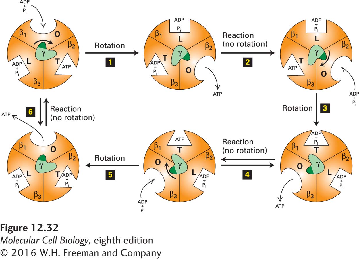
FIGURE 12-32 The binding-change mechanism of ATP synthesis from ADP and Pi. This view is looking up at F1 from the membrane surface (see Figure 12-31). As the γ subunit rotates by 120° in the center, each of the otherwise identical F1 β subunits alternates between three conformational states (O, open, with oval representation of the binding site; L, loose, with a rectangular binding site; T, tight, with a triangular site) that differ in their binding affinities for ATP, ADP, and Pi. The cycle begins (upper left) when ADP and Pi bind loosely to one of the three β subunits (here, arbitrarily designated β1) whose nucleotide-binding site is in the O (open) conformation. Proton flux through the F0 portion of the protein powers a 120° rotation of the γ subunit (relative to the fixed β subunits) (step 1). This causes the rotating γ subunit, which is asymmetric, to push differentially against the β subunits, resulting in a conformational change and an increase in the binding affinity of the β1 subunit for ADP and Pi (O → L), an increase in the binding affinity of the β3 subunit for ADP and Pi that were previously bound (L → T), and a decrease in the binding affinity of the β2 subunit for a previously bound ATP (T → O), causing release of the bound ATP. Step 2: Without additional rotation, the ADP and Pi in the T site (here, in the β3 subunit) form ATP, a reaction that does not require an input of additional energy due to the special environment in the active site of the T state. At the same time, a new ADP and Pi bind loosely to the unoccupied O site on β2. Step 3: Proton flux powers another 120° rotation of the γ subunit, consequent conformational changes in the binding sites (L → T, O → L, T → O), and release of ATP from β3. Step 4: Without additional rotation, the ADP and Pi in the T site of β1 form ATP, and additional ADP and Pi bind to the unoccupied O site on β3. The process continues with rotation (step 5) and ATP formation (step 6) until the cycle is complete, with three ATPs having been produced for every 360° rotation of γ. See P. Boyer, 1989, FASEB J. 3:2164; Y. Zhou et al., 1997, Proc. Natl. Acad. Sci. USA 94:10583; and M. Yoshida, E. Muneyuki, and T. Hisabori, 2001, Nat. Rev. Mol. Cell Biol. 2:669.