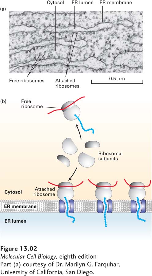
FIGURE 13- 2 Structure of the rough ER. (a) Electron micrograph of ribosomes attached to the rough ER in a pancreatic acinar cell. Most of the proteins synthesized by this type of cell are secretory proteins and are formed on membrane- attached ribosomes. A few unattached (free) ribosomes are evident; presumably, these ribosomes are synthesizing cytosolic or other nonsecretory proteins. (b) Schematic representation of protein synthesis on the ER. Note that membrane- bound and free cytosolic ribosomes are identical. Membrane- bound ribosomes are recruited to the ER during synthesis of a polypeptide containing an ER signal sequence.
[Part (a) courtesy of Dr. Marilyn G. Farquhar, University of California, San Diego.]
[Leave] [Close]