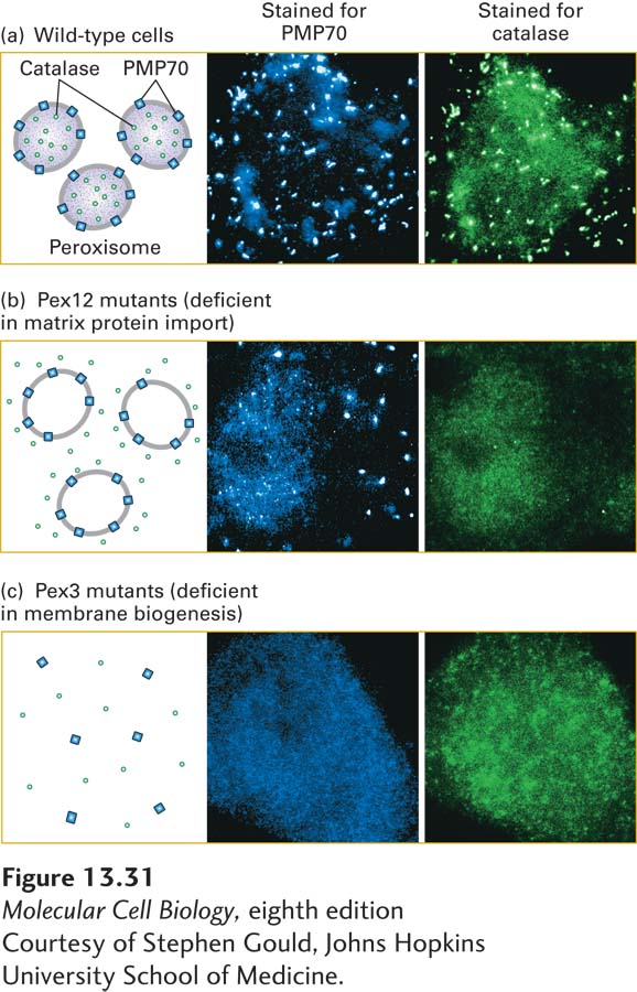
EXPERIMENTAL FIGURE 13- 31 Studies reveal different pathways for incorporation of peroxisomal membrane and matrix proteins. Cells were stained with fluorescent antibodies to PMP70, a peroxisomal membrane protein, or with fluorescent antibodies to catalase, a peroxisomal matrix protein, then viewed in a fluorescence microscope. (a) In wild- type cells, both peroxisomal membrane and matrix proteins are visible as bright foci in numerous peroxisomal bodies. (b) In cells from a Pex12- deficient patient, catalase is distributed uniformly throughout the cytosol, whereas PMP70 is localized normally to peroxisomal bodies. (c) In cells from a Pex3- deficient patient, peroxisomal membranes cannot assemble, and as a consequence, peroxisomal bodies do not form. Thus both catalase and PMP70 are mis- localized to the cytosol.
[Courtesy of Stephen Gould, Johns Hopkins University School of Medicine.]
[Leave] [Close]