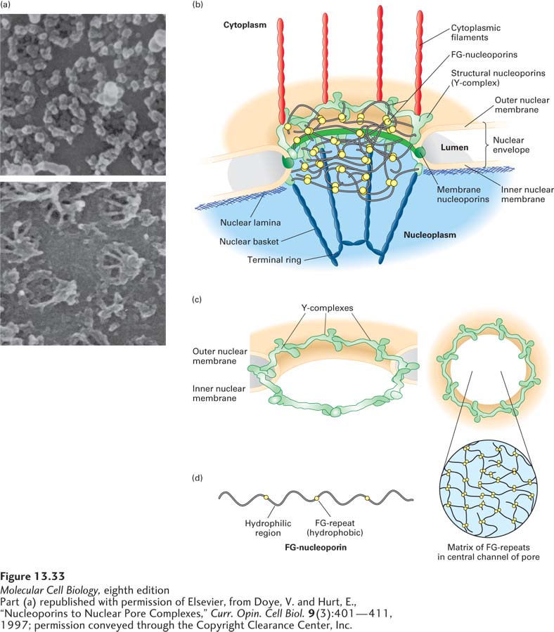
FIGURE 13- 33 Nuclear pore complex at different levels of resolution. (a) Nuclear envelopes from the large nuclei of Xenopus oocytes, visualized by scanning electron microscopy. Top: View of the cytoplasmic face reveals the octagonal shape of the membrane- embedded portion of nuclear pore complexes. Bottom: View of the nucleoplasmic face shows the nuclear basket that extends from the membrane- embedded portion. (b) Cutaway model of the nuclear pore complex, showing the major structural features formed by membrane nucleoporins, structural nucleoporins, and FG- nucleoporins. (c) Sixteen copies of the Y- complex form a major part of the structural scaffold of the nuclear pore complex. The three- dimensional structure of the Y- complex is modeled into the pore structure. Note the twofold symmetry across the double membrane of the nucleus (left) and the eightfold rotational symmetry around the axis of the pore (right). (d) The FG- nucleoporins have extended disordered structures that are composed of repeats of the sequence Phe– Gly interspersed with hydrophilic regions (left). The FG- nucleoporins are most abundant in the central part of the pore, and the FG- repeat sequences are thought to fill the central channel with a gel- like matrix (right). See K. Ribbeck and D. Görlich, 2001, EMBO J. 20:1320– 1330 and M. P. Rout and J. D. Atchison, 2001, J. Biol. Chem. 276:16593.
[Part (a) republished with permission of Elsevier, from Doye, V. and Hurt, E., “Nucleoporins to Nuclear Pore Complexes,” Curr. Opin. Cell Biol. 9(3):401– 411, 1997; permission conveyed through the Copyright Clearance Center, Inc.]
[Leave] [Close]