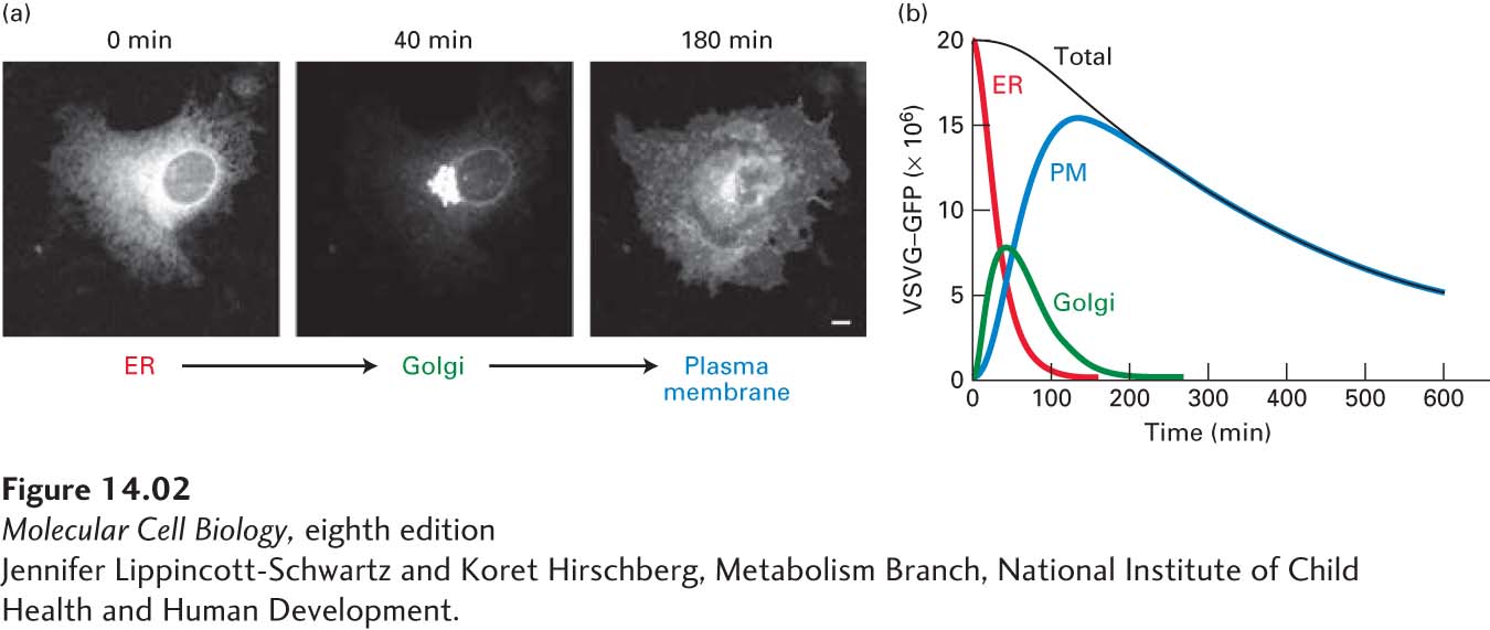
EXPERIMENTAL FIGURE 14- 2 Protein transport through the secretory pathway can be visualized by fluorescence microscopy of cells producing a GFP- tagged membrane protein. Cultured cells were transfected with a hybrid gene encoding the viral membrane glycoprotein VSV G linked to the gene for green fluorescent protein (GFP). A temperature- sensitive mutant version of the viral gene was used so that newly made hybrid protein (VSVG- GFP) was retained in the ER at 40 °C, but was released for transport at 32 °C. (a) Fluorescence micrographs of cells just before and at two times after they were shifted to the lower temperature. Movement of VSVG- GFP from the ER to the Golgi and finally to the cell surface occurred within 180 minutes. The scale bar is 5 µm. (b) Plot of the amount of VSVG- GFP in the endoplasmic reticulum (ER), Golgi, and plasma membrane (PM) at different times after the shift to the permissive temperature. The kinetics of transport from one organelle to another can be reconstructed from computer analysis of these data. The decrease in total fluorescence that occurs at later times probably results from slow inactivation of GFP fluorescence.
[Jennifer Lippincott- Schwartz and Koret Hirschberg, Metabolism Branch, National Institute of Child Health and Human Development.]
[Leave] [Close]