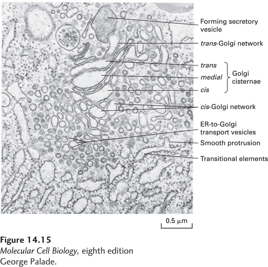
EXPERIMENTAL FIGURE 14- 15 Electron micrograph of the Golgi complex in an pancreatic acinar cell reveals secretory and retrograde transport vesicles. A large secretory vesicle can be seen forming from the trans-Golgi network. Elements of the rough ER are on the bottom and left in this micrograph. Adjacent to the rough ER are transitional elements from which smooth protrusions appear to be budding. These buds form the small vesicles that transport secretory proteins from the rough ER to the Golgi complex. Interspersed among the Golgi cisternae are other small vesicles now known to function in retrograde, not anterograde, transport.
[George Palade.]
[Leave] [Close]