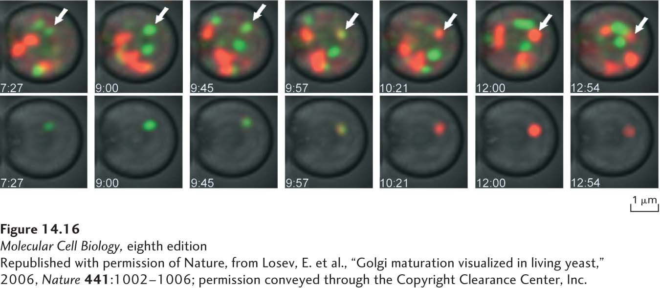
EXPERIMENTAL FIGURE 14- 16 Fluorescence- tagged fusion proteins demonstrate Golgi cisternal maturation in a live yeast cell. Yeast cells expressing the early Golgi protein Vrg4 fused to GFP (green fluorescence) and the late Golgi protein Sec7 fused to DsRed (red fluorescence) were imaged by time- lapse microscopy. The top series of images, taken approximately 1 minute apart, shows a collection of Golgi cisternae, which at any one time are labeled with either Vrg4 or Sec7. The bottom series of images show just one Golgi cisterna, isolated by digital processing of the image. First only Vrg4- GFP is located in the isolated cisterna, and later only Sec7- DsRed is located in the isolated cisterna, following a brief period in which both proteins are co- localized in this compartment. This experiment is a direct demonstration of the cisternal maturation hypothesis, showing that the composition of individual cisternae follows a process of maturation characterized by loss of early Golgi proteins and gain of late Golgi proteins.
[Republished with permission of Nature, from Losev, E. et al., “Golgi maturation visualized in living yeast,” 2006, Nature 441:1002– 1006; permission conveyed through the Copyright Clearance Center, Inc.]
[Leave] [Close]