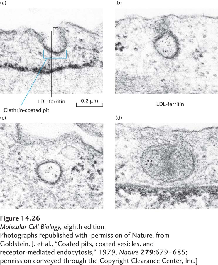
EXPERIMENTAL FIGURE 14- 26 The initial stages of receptor- mediated endocytosis of low- density lipoprotein (LDL) particles are revealed by electron microscopy. Cultured human fibroblasts were incubated in a medium containing LDL particles covalently linked to the electron- dense, iron- containing protein ferritin; each small iron particle in ferritin is visible as a small dot under the electron microscope. Cells were initially incubated at 4 °C; at this temperature LDL can bind to its receptor, but internalization does not occur. After excess LDL not bound to the cells was washed away, the cells were warmed to 37 °C and then prepared for microscopy at periodic intervals. (a) A coated pit, showing the clathrin coat on the inner (cytosolic) surface of the pit, soon after the temperature was raised. (b) A pit containing LDL apparently closing on itself to form a coated vesicle. (c) A coated vesicle containing ferritin- tagged LDL particles. (d) Ferritin- tagged LDL particles in a smooth- surfaced early endosome 6 minutes after internalization began. See also M. S. Brown and J. Goldstein, 1986, Science 232:34.
[Photographs republished with permission of Nature, from Goldstein, J. et al., “Coated pits, coated vesicles, and receptor- mediated endocytosis,” 1979, Nature 279:679– 685; permission conveyed through the Copyright Clearance Center, Inc.]
[Leave] [Close]