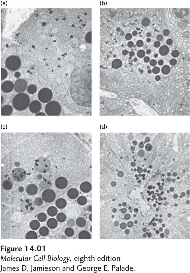
FIGURE 1 The synthesis and movement of guinea pig pancreatic secretory proteins as revealed by electron microscopic autoradiography. After a period of labeling with [3H]leucine, guinea pig pancreatic tissue was fixed, sectioned for electron microscopy, and subjected to autoradiography. The radioactive decay of [3H] in newly synthesized proteins produces autoradiographic grains in an emulsion placed over the cell section (which appear in the micrograph as dense, wormlike granules); these grains mark the positions of newly synthesized proteins. (a) At the end of a 3- 7- 7- 7-
[James D. Jamieson and George E. Palade.]