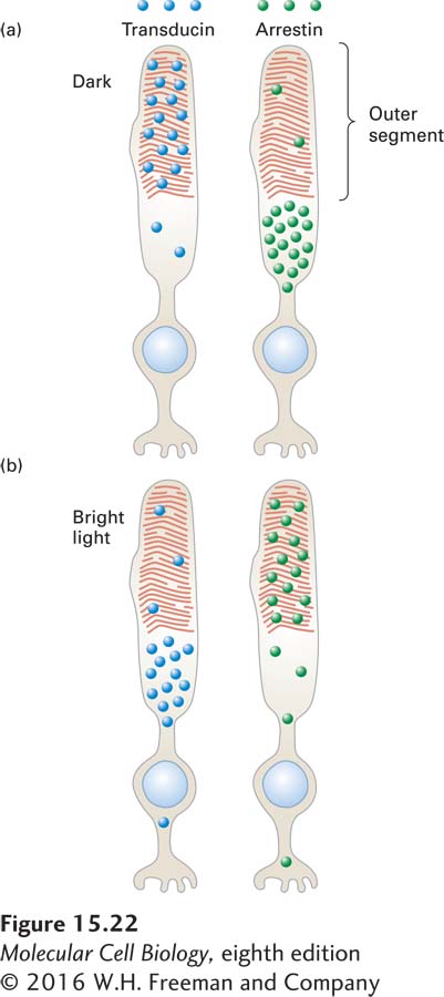
FIGURE 15- 22 Schematic illustration of transducin and arrestin distribution in dark- adapted and light- adapted rod cells. (a) In the dark, most transducin (blue circles) is localized to the outer segment, while most arrestin (green circles) is found in other parts of the cell; in this condition, vision is most sensitive to very low light levels. (b) In bright light, little transducin is found in the outer segment, and abundant arrestin is found there; in this condition, vision is relatively insensitive to small changes in light. The coordinated movement of these proteins contributes to our ability to perceive images over a 100,000- fold range of ambient light levels. See P. Calvert et al., 2006, Trends Cell Biol. 16:560.
[Leave] [Close]