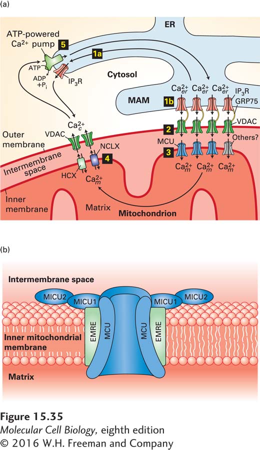
FIGURE 15- 35 Movement of Ca2+ between the cytosol, mitochondrion, and endoplasmic reticulum. (a) The ER is the main intracellular storage depot for Ca2+. Binding of IP3 to IP3-gated Ca2+ channels (IP3R) in the membrane of the endoplasmic reticulum releases Ca2+ into the cytosol (step 1a); binding also opens IP3-gated Ca2+ channels in the mitochondria- associated membranes (MAMs) of the ER (step 1b). Step 2: VDAC channels in the outer mitochondrial membrane adjacent to MAMs are physically linked to IP3Rs by the GRP75 protein; they efficiently pass the Ca2+ released from the MAMs into the intermembrane space. Step 3: The high concentration of Ca2+ in the intermembrane space induces the opening of MCUs or other Ca2+ channels in the inner membrane, resulting in the flow of Ca2+ into the mitochondrial matrix. Step 4: Over time, Ca2+ is released from the mitochondria by Ca2+/Na+ (NCLX) and Ca2+/H+ (HCX) antiporters in the inner membrane, then transferred into the cytosol through VDAC or other Ca2+ channels in the outer membrane. Finally, pumping of Ca2+ from the cytosol by ATP- powered Ca2+ pumps in the ER membrane (step 5) or plasma membrane restores the high ER Ca2+ and low cytosolic Ca2+ levels. (b) Model of the mitochondrial calcium uniporter complex. A multimer of MCU subunits forms the regulated Ca2+ pore. Additional subunits include the integral membrane protein EMRE and regulatory subunits MICU1 and MICU2. Ca2+ binding to the MICU subunits opens the MCU pore, resulting in the flow of the Ca2+ from the intermembrane space into the matrix. See M. Schäfer et al., 2014, Cell Tissue Res. 357:395 and K. Kamer and V. Mootha, 2015, Nat. Rev. Mol. Cell Biol. 16:545.
[Leave] [Close]