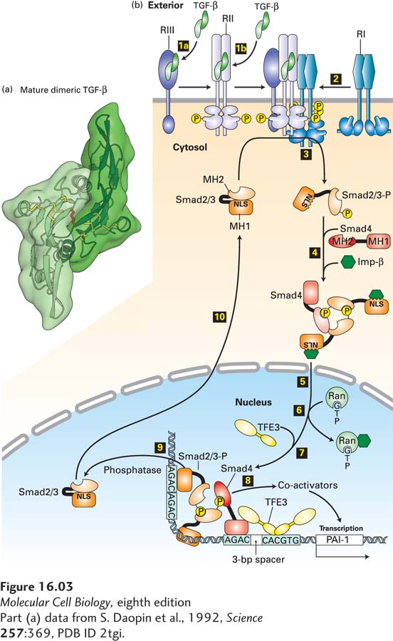
FIGURE 16- 3 TGF- β/Smad signaling pathway. (a) Ribbon diagram structure of a mature TGF- β dimer. The three intrachain disulfide linkages (yellow) in each monomer form a cystine- knot domain; another disulfide bond (red) links the two monomers. (b) Step 1a: In some cells, TGF- β binds to the type III TGF- β receptor (RIII), which presents TGF- β to the type II receptor (RII). Step 1b: In other cells, TGF- β binds directly to RII, a constitutively active kinase. Step 2: Ligand- bound RII recruits and phosphorylates the juxtamembrane segment of the type I TGF- β receptor (RI), which does not directly bind TGF- β. This releases the inhibition of RI kinase activity. Step 3: Activated RI then phosphorylates Smad2 or Smad3 (shown here as Smad2/3), causing a conformational change that unmasks its nuclear- localization signal (NLS). Step 4: Two phosphorylated molecules of Smad2/3 bind to a co- Smad (Smad4) molecule, which is not phosphorylated, and to an importin, forming a large cytosolic complex. Steps 5 and 6: After the entire complex translocates into the nucleus, Ran·GTP causes dissociation of the importin, as discussed in Chapter 13. Step 7: A nuclear transcription factor (e.g., TFE3) then associates with the Smad2/3/Smad4 complex, forming an activation complex that cooperatively binds to regulatory sequences of a target gene. Step 8: This complex then recruits transcriptional co- activators and induces gene transcription (see Chapter 9). Smad2/3 is dephosphorylated by a nuclear phosphatase (step 9) and recycles through a nuclear pore to the cytosol (step 10), where it can be reactivated by another TGF- β receptor complex. Shown at the bottom is the activation complex for the gene encoding plasminogen activator inhibitor (PAI- 1); similar transcription complexes activate expression of genes encoding other extracellular- matrix proteins such as fibronectin. See A. Moustakas and C.-H. Heldin, 2009, Development 136:3699, and D. Clarke and X. Liu, 2008, Trends Cell Biol. 18:430.
[Part (a) data from S. Daopin et al., 1992, Science 257:369, PDB ID 2tgi.]
[Leave] [Close]