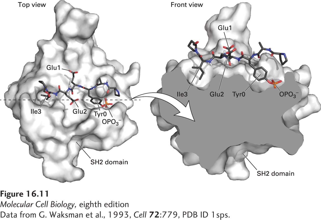
FIGURE 16- 11 Surface model of an SH2 domain bound to a phosphotyrosine- containing peptide. The peptide bound by this SH2 domain from Src tyrosine kinase (blue backbone with red oxygen atoms) is shown in stick form. The SH2 domain binds strongly to short target peptides containing a critical four- residue core sequence: phosphotyrosine (Tyr0 and OPO3−)–glutamic acid (Glu1)–glutamic acid (Glu2)–isoleucine (Ile3). Binding resembles the insertion of a two- pronged “plug”—the phosphotyrosine and isoleucine side chains of the peptide— into a two- pronged “socket” in the SH2 domain. The two glutamate residues are bound to sites on the surface of the SH2 domain between the two sockets.
[Data from G. Waksman et al., 1993, Cell 72:779, PDB ID 1sps.]
[Leave] [Close]