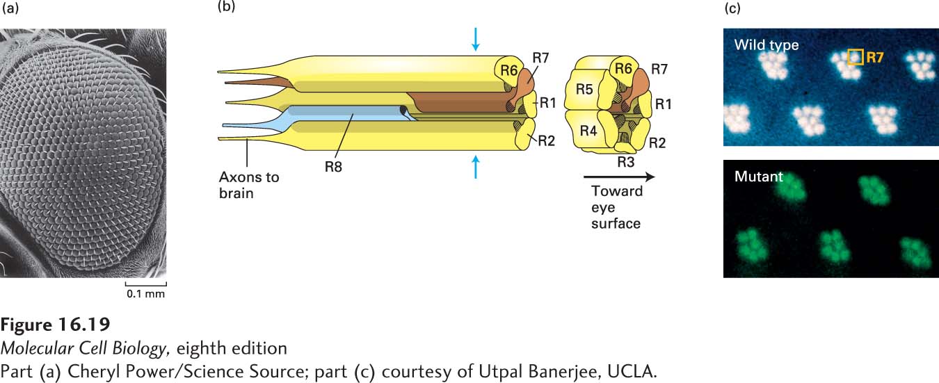
FIGURE 16- 19 The compound eye of Drosophila melanogaster. (a) Scanning electron micrograph showing the individual ommatidia that compose the fruit fly eye. (b) Longitudinal and cutaway views of a single ommatidium. Each of these tubular structures contains eight photoreceptors, designated R1– R8, which are long, cylindrically shaped light- sensitive cells. R1– R6 (yellow) extend throughout the depth of the retina, whereas R7 (brown) is located toward the surface of the eye and R8 (blue) toward the back side, where the axons exit. (c) Comparison of eyes from wild- type and sevenless mutant flies viewed by a special technique that can distinguish the photoreceptors in an ommatidium. The plane of sectioning is indicated by the blue arrows in (b), and the R8 cell is out of the plane of these images. The seven photoreceptors in this plane are easily seen in the wild- type ommatidia (top), whereas only six are visible in the mutant ommatidia (bottom). The eyes of flies with the sevenless mutation lack the R7 cell.
[Part (a) Cheryl Power/Science Source; part (c) courtesy of Utpal Banerjee, UCLA.]
[Leave] [Close]