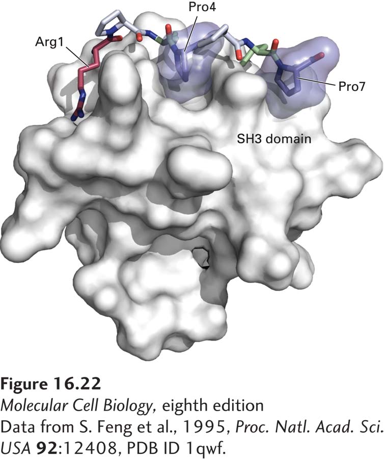
FIGURE 16- 22 Surface model of an SH3 domain bound to a target peptide. The short, proline- rich target peptide is shown as a space- filling model. In this target peptide, two prolines (Pro4 and Pro7, dark blue) fit into binding pockets on the surface of the SH3 domain. Interactions involving an arginine (Arg1, red), two other prolines (gray), and other residues in the target peptide (green) determine the specificity of binding.
[Data from S. Feng et al., 1995, Proc. Natl. Acad. Sci. USA 92:12408, PDB ID 1qwf.]
[Leave] [Close]