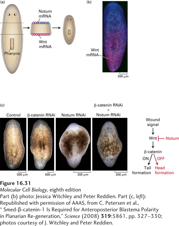
FIGURE 16- 31 Gradients of Wnt are essential for normal regeneration of a head and a tail by planaria. (a) As indicated in the diagram, a small piece excised from the middle of the body of the planarian S. mediterranea was placed in culture. In situ mRNA hybridization performed after 4 days indicated that Wnt mRNA (pink dots) was expressed in cells at both the anterior and posterior wound sites, but that the Wnt inhibitor Notum (blue dots) was expressed only at the anterior wound site. Thus a posterior- to- anterior gradient of Wnt protein is formed. (b) As shown by in situ hybridization, the WntP- 2 gene (pink dots) is expressed in a posterior- to- anterior gradient in adult planaria. See J. Witchley et al., 2013, Cell Rep. 4:633. (c) After 14 days, a normal, albeit smaller, worm has regenerated a head, easily visualized by the two eyes, from the anterior wound site and a tail from the posterior. Treatment of the excised body piece with an inhibitory RNA specific for β-catenin results in regeneration of a two- headed planarian, whereas treatment with an inhibitory RNA specific for Notum results in regeneration of a two- tailed planarian. Treatment of the excised body piece with two inhibitory RNAs, one specific for β-catenin and the other for Notum, results in a phenotype similar to that caused by the loss of β-catenin alone: a two- headed planarian is regenerated. These experiments give rise to the model depicted in part (c), in which β-catenin, stabilized by addition of Wnt to cells, causes expression of genes that promote tail formation; inhibition of Wnt/β-catenin signaling by Notum causes a head to be formed.
[Part (b) photo: Jessica Witchley and Peter Reddien. Part (c, left): Republished with permission of AAAS, from C. Petersen et al., “Smed- β-catenin- 1 Is Required for Anteroposterior Blastema Polarity in Planarian Regeneration,” Science (2008) 319:5861, pp. 327- 330; photos courtesy of J. Witchley and Peter Reddien.]
[Leave] [Close]