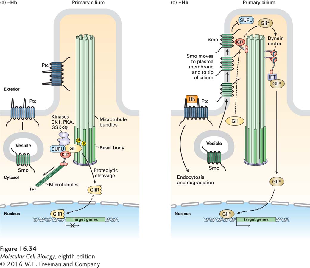
FIGURE 16- 34 Hedgehog signaling in vertebrates. Hedgehog (Hh) signaling occurs in primary cilia, but otherwise the overall process is similar to that in Drosophila. (a) In the absence of Hh, Ptc is localized to the ciliary membrane and the base of the cilium. In an unknown manner, Ptc blocks the entry of Smo to the plasma membrane; Smo is present mainly in the membrane of internal vesicles. The kinesin Kif7 (the Cos2 homolog) binds to microtubules at the cilium base, where it prevents the transcription factor Gli (the vertebrate homolog of Ci) from entering the cilium. Kif7 and Gli are part of a complex that includes SUFU and the kinases CK1, PKA, and GSK3β, which phosphorylate Gli and promote its proteolytic cleavage to form the repressor GliR. (b) Hh binding triggers endocytosis and degradation of the Hh/Ptc complex, movement of Smo to the plasma membrane, and then, together with several proteins bound to it, its movement to the tip of the cilium. There Smo triggers dissociation of the SUFU- Gli complex. Rather than being degraded, Gli accumulates, becomes modified by addition of several phosphate and acetyl groups, forming the active Gli*, and is then transported down the cilium by a dynein motor protein. Gli* is then released into the cytosol, translocates into the nucleus, and activates gene expression. See J. Briscoe and P. Thérond, 2013, Nat. Rev. Mol. Cell Biol. 14:416.
[Leave] [Close]