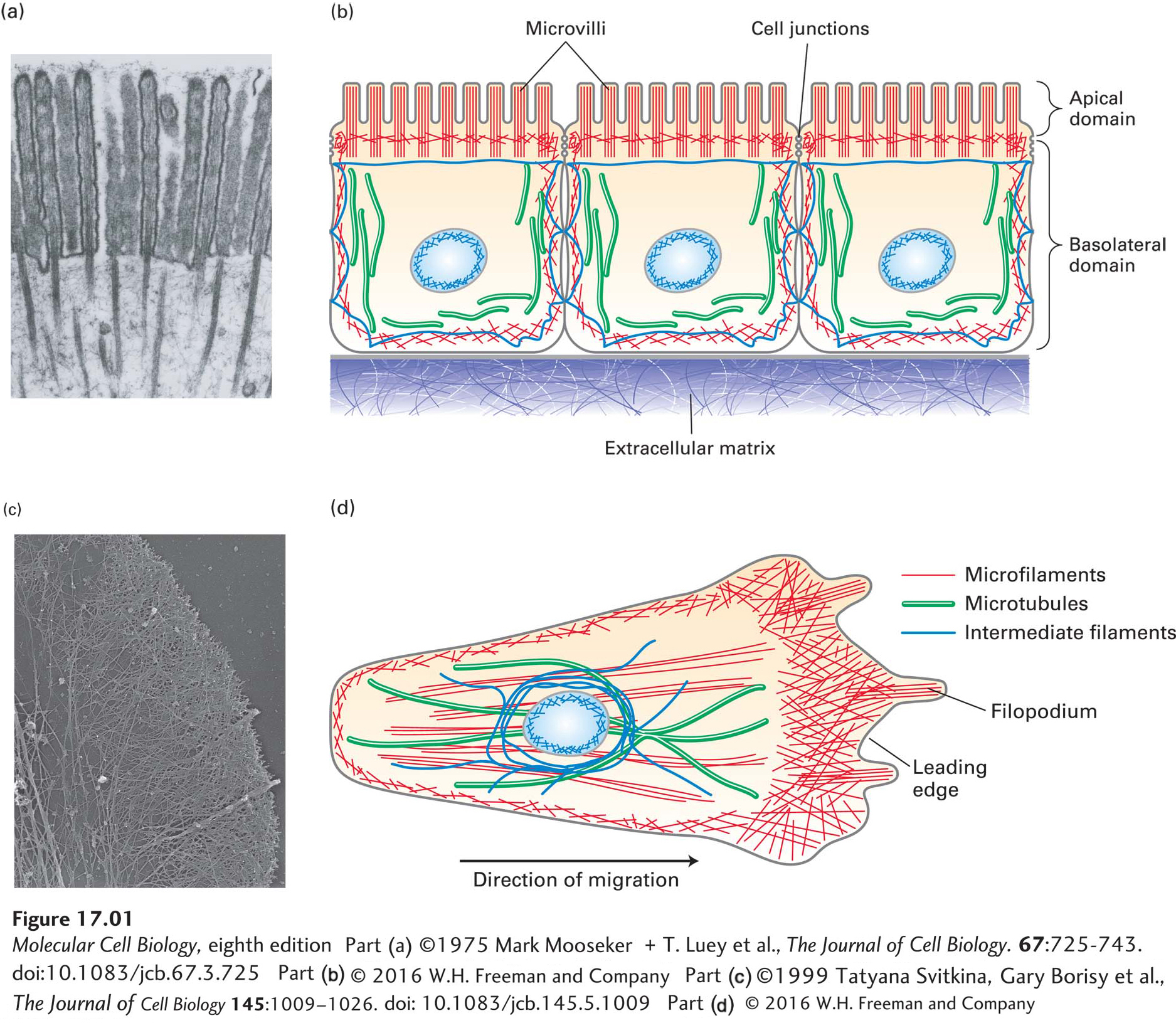
FIGURE 17- 1 Overview of the cytoskeletons of an epithelial cell and a migrating cell. (a) Transmission electron micrograph of a thin section of an epithelial cell from the small intestine, showing the core bundles of microfilaments that provide support to the microvilli. (b) Epithelial cells are highly polarized, with distinct apical and basolateral domains. An intestinal epithelial cell transports nutrients into the cell through the apical domain and out of the cell across the basolateral domain. (c) Transmission electron micrograph of part of the leading edge of a migrating cell. The cell was treated with a mild detergent to dissolve the membranes, which also allows solubilization of most cytoplasmic components. The remaining cytoskeleton was shadowed with platinum and visualized in the electron microscope. Note the network of actin filaments visible in this micrograph. (d) A migrating cell, such as a fibroblast or a macrophage, has morphologically distinct domains, with a leading edge at the front. Microfilaments are indicated in red, microtubules in green, and intermediate filaments in dark blue. The position of the nucleus (light blue oval) is also shown.
[Part (a) ©1975 Mark Mooseker + T. Luey et al., The Journal of Cell Biology. 67:725- 743. doi:10.1083/jcb.67.3.725 Part (c) ©1999 Tatyana Svitkina, Gary Borisy et al., The Journal of Cell Biology 145:1009– 1026. doi: 10.1083/jcb.145.5.1009]
[Leave] [Close]