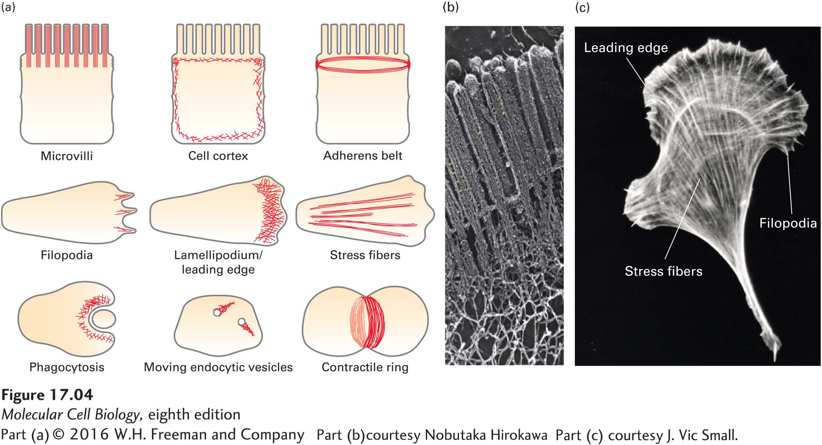
FIGURE 17- 4 Examples of microfilament- based structures. (a) In each panel, microfilaments are depicted in red. (b) The apical region of a polarized epithelial cell, showing the bundles of actin filaments that make up the cores of the microvilli. The sample was prepared using a rapid freeze, deep etch, rotary shadow protocol and viewed by transmission electron microscopy. (c) A cell moving toward the top of the page, stained for actin with fluorescent phalloidin, a drug that specifically binds F- actin. Note how many different organizations of microfilaments can exist in one cell.
[Part (b) courtesy Nobutaka Hirokawa; Part (c) courtesy J. Vic Small.]
[Leave] [Close]