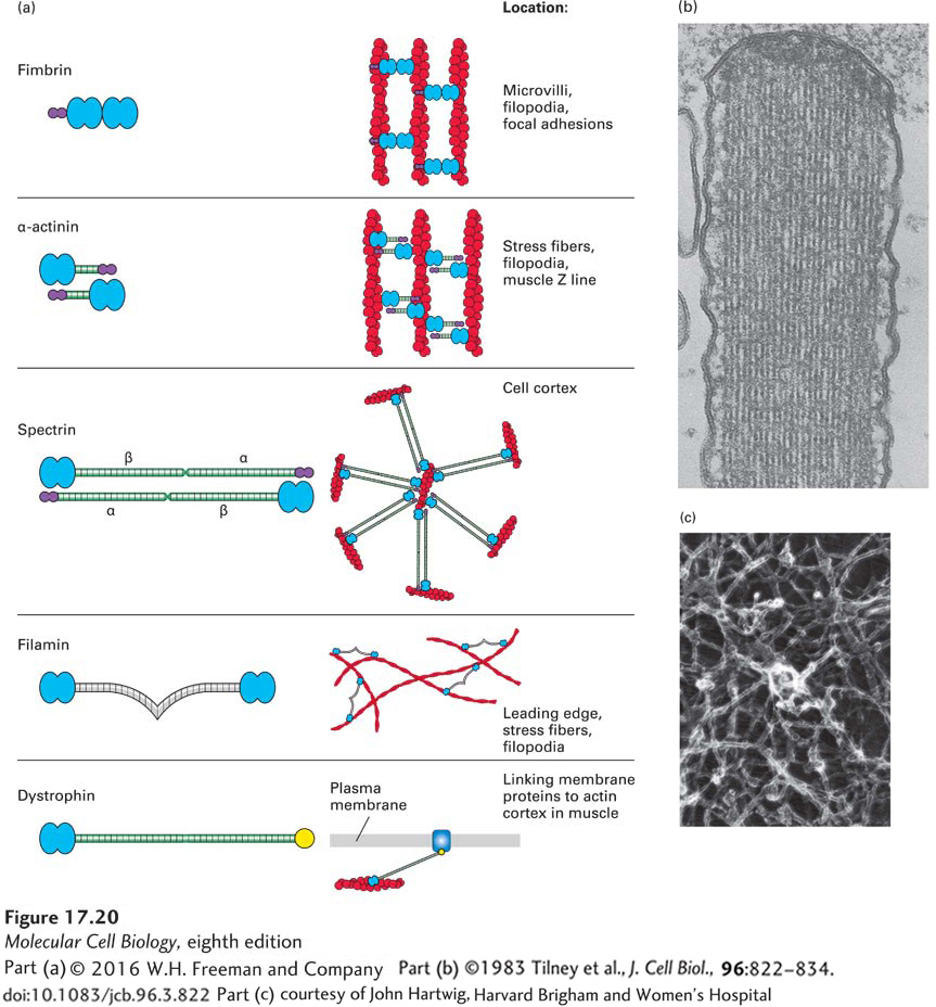
FIGURE 17- 20 Actin cross- linking proteins. Actin cross- linking proteins mold F- actin filaments into diverse structures. (a) Examples of four F- actin cross- linking proteins, all of which have two domains (blue) that bind F- actin. Some have a Ca2+-binding site (purple) that inhibits their activity at high levels of free Ca2+. Also shown is dystrophin, which has an actin- binding site on its N- terminal end and a C- terminal domain that binds the membrane protein dystroglycan. (b) Transmission electron micrograph of a thin section of a stereocilium (an unfortunate name, since it is really a giant microvillus) on a sensory hair cell in the inner ear. This structure contains a bundle of actin filaments cross- linked by fimbrin, a small cross- linking protein that allows for close and regular interaction of actin filaments. (c) Long cross- linking proteins such as filamin are flexible and can thus cross- link actin filaments into a loose network.
[Part (b) ©1983 Tilney et al., J. Cell Biol., 96:822– 834. doi: 10.1083/jcb.96.3.822; part (c) courtesy of John Hartwig, Harvard Brigham and Women’s Hospital.]
[Leave] [Close]