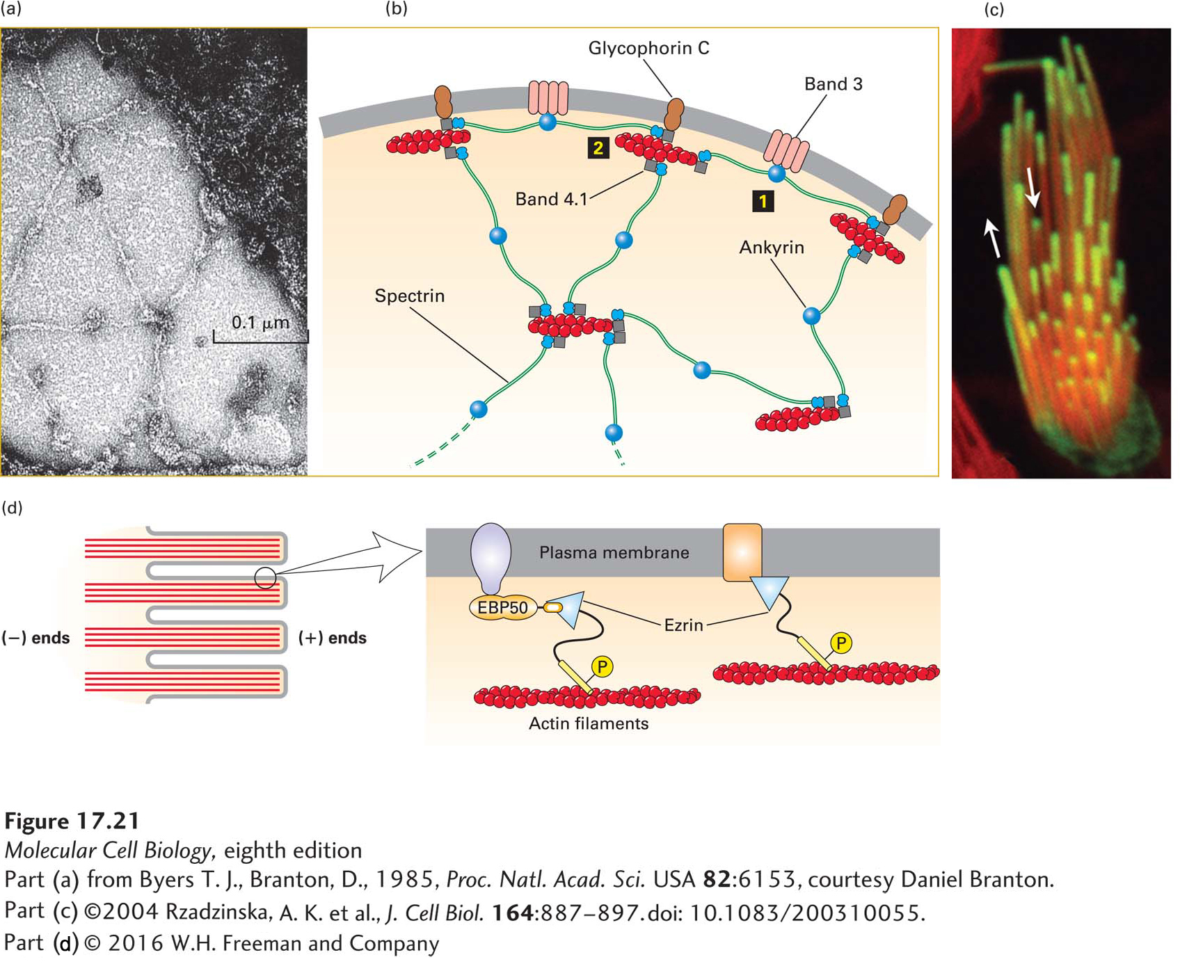
FIGURE 17- 21 Lateral attachment of microfilaments to membranes. (a) Electron micrograph of the erythrocyte membrane showing the spoke- and- hub organization of the cortical cytoskeleton supporting the plasma membrane in human erythrocytes. The long spokes are composed mainly of spectrin and can be seen to intersect at the hubs, or membrane- attachment sites. The darker spots along the spokes are ankyrin molecules, which link spectrin to integral membrane proteins. (b) Diagram of the erythrocyte cytoskeleton, showing the two main types of membrane attachments: 1 through ankyrin to band 3 and 2 through band 4.1 to glycophorin C. (c) Actin is incorporated into the tips of stereocilia (giant microvilli). Cells with stereocilia were transfected to express GFP- labeled actin for a short period of time and then counterstained with rhodamine- phalloidin to stain all the F- actin. The experiment shows that new actin is incorporated at the tips of the stereocilia. (d) Ezrin, a member of the ezrin- radixin- moesin (ERM) family, links actin filaments laterally to the plasma membrane in surface structures such as microvilli; attachment can be direct or indirect. Ezrin, activated by phosphorylation (P), links directly to the cytoplasmic region of transmembrane proteins (right) or indirectly through a scaffolding protein such as EBP50 (left). See R. G. Fehon et al., 2010, Nature Rev. Mol. Cell Biol. 11:276, S. E. Lux, 1979, Nature 281:426, and E. J. Luna and A. L. Hitt, 1992, Science 258:955.
[Part (a) from Byers T. J., Branton, D., 1985, Proc. Natl. Acad. Sci. USA 82:6153, courtesy Daniel Branton; part (c) ©2004 Rzadzinska, A. K. et al., J. Cell Biol. 164:887– 897. doi: 10.1083/200310055.]
[Leave] [Close]