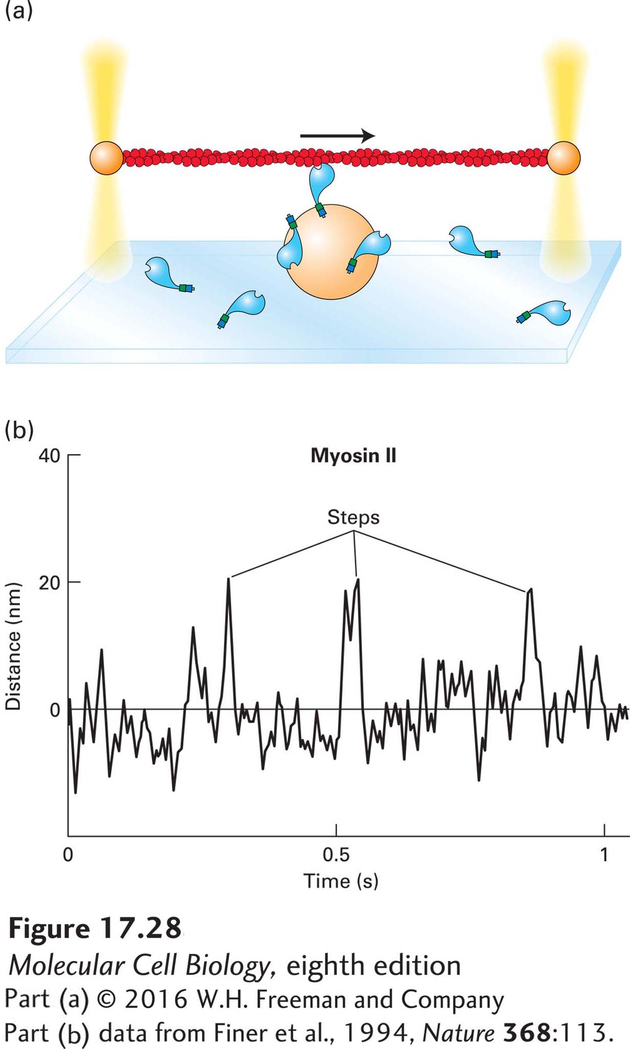
EXPERIMENTAL FIGURE 17- 28 Measuring myosin step size and force with actin held by optical traps. (a) Optical trap techniques can be used to determine the step size and force generated by a single myosin molecule. In an optical trap, the beam of an infrared laser is focused by a light microscope on a latex bead (or any other object that does not absorb infrared light) to capture and hold the bead in the center of the beam. The strength of the force holding the bead is adjusted by increasing or decreasing the intensity of the laser beam. In this experiment, an actin filament is held between two optical traps. The actin filament is then lowered onto a third bead coated with a dilute concentration of myosin molecules. If the actin filament encounters a myosin molecule in the presence of ATP, the myosin will pull on the actin filament, which allows the investigators to measure both the force generated and the step size the myosin takes. (b) Using an optical trap setup, investigators have analyzed the behavior of myosin II. As shown by the peaks in the trace, myosin II takes erratic small steps (5– 15 nm), which means that it binds the actin filament, moves, and then lets go. It is therefore a nonprocessive motor.
[Part (b) data from Finer et al., 1994, Nature 368:113.]
[Leave] [Close]