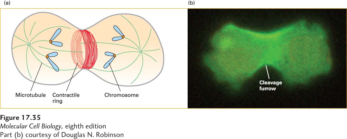
EXPERIMENTAL FIGURE 17- 35 Fluorescent antibodies reveal the localization of myosin I and myosin II during cytokinesis. (a) Diagram of a cell going through cytokinesis, showing the mitotic spindle (microtubules green, chromosomes blue) and the contractile ring with actin filaments (red). (b) Fluorescence micrograph of a Dictyostelium amoeba expressing GFP- myosin- II reveals myosin- II enrichment in the cleavage furrow cortex, as the cell pinches into two during cytokinesis.
[Part (b) courtesy of Douglas N. Robinson]
[Leave] [Close]