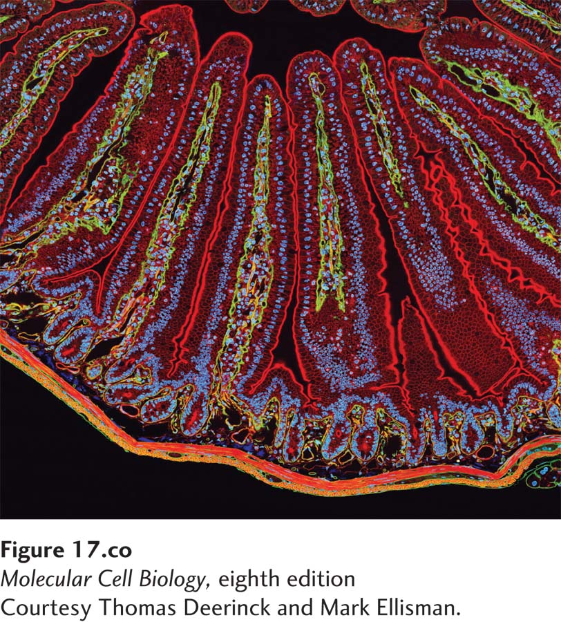
A section of mouse intestine stained for actin (red), the extracellular matrix protein laminin (green), and DNA (blue). Each blue dot of DNA indicates the presence of a cell. Actin in the microvilli on the apical end of the epithelial cells can be seen lining the surface facing the lumen (top). Actin can also be seen prominently in the smooth muscle that surrounds the intestine (bottom).
[Courtesy Thomas Deerinck and Mark Ellisman.]
[Leave] [Close]