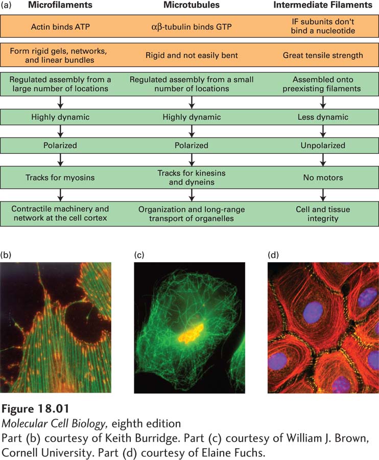
image
FIGURE 18-1 Overview of the physical properties and functions of the three filament systems in animal cells. (a) Biophysical and biochemical properties (orange) and biological properties (green) are shown for each filament type. The micrographs (b–d) show examples of each filament type in a particular cellular context, but note that microtubules also make up other structures, and that intermediate filaments also line the inner surface of the nucleus. (b) Cultured cells stained for actin (green) and sites of actin attachment to the substratum (orange). (c) Localization of microtubules (green) and the Golgi complex (yellow). Notice the central location of the Golgi complex, which is collected there by transport along microtubules. (d) Localization of cytokeratins (red), a type of intermediate filament, and a component of desmosomes (yellow) in epithelial cells. Cytokeratins from individual cells are attached to each other through the desmosomes.
[Part (b) courtesy of Keith Burridge. Part (c) courtesy of William J. Brown, Cornell University. Part (d) courtesy of Elaine Fuchs.]