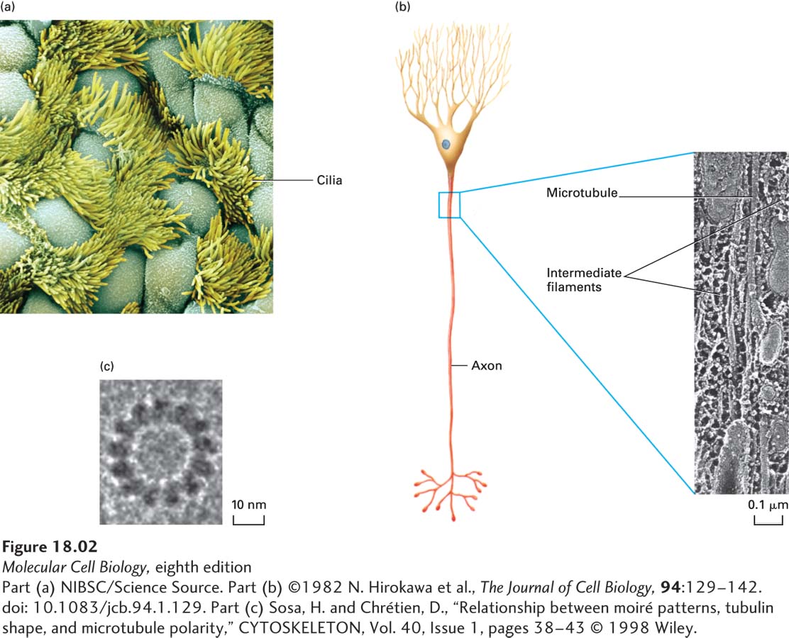
image
FIGURE 18-2 Microtubules are found in many different locations, and all have similar structures. (a) Surface of the ciliated epithelium lining a rabbit oviduct viewed in a scanning electron microscope. Beating cilia, each of which has a core of microtubules, propel eggs down the oviduct. (b) Microtubules and intermediate filaments in a quick-frozen and deep-etched frog axon visualized in a transmission electron microscope. (c) High-magnification view of a single microtubule showing the 13 repeating units known as protofilaments.
[Part (a) NIBSC/Science Source. Part (b) ©1982 N. Hirokawa et al., The Journal of Cell Biology, 94:129–142. doi: 10.1083/jcb.94.1.129. Part (c) Sosa, H. and Chrétien, D., “Relationship between moiré patterns, tubulin shape, and microtubule polarity,” CYTOSKELETON, Vol. 40, Issue 1, pages 38-43 © 1998 Wiley.]