image
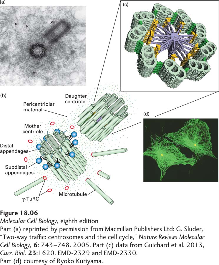
FIGURE 18-6 Structure of centrosomes. (a) Thin section of an animal-cell centrosome showing the two centrioles at right angles to each other, surrounded by pericentriolar material (arrows). (b) Diagram of a centrosome showing the mother and daughter centrioles, each of which consists of nine linked triplet microtubules, embedded in pericentriolar material that contains γ-TuRC nucleating structures. The mother centriole is distinct from the daughter as it has distal appendages (blue spheres). (c) A tomographic image of a section through a daughter centriole of the alga Chlamydomonas. The daughter centriole is believed to be templated by a nine-fold symmetric cartwheel structure that is later removed. (d) Immunofluorescence micrograph showing the microtubule array (green) in a cultured animal cell and the location of the MTOC, using an antibody to a centrosomal protein (yellow).
[Part (a) reprinted by permission from Macmillan Publishers Ltd: G. Sluder, “Two-way traffic: centrosomes and the cell cycle,” Nature Reviews Molecular Cell Biology, 6: 743-748. 2005. Part (c) data from Guichard et al. 2013, Curr. Biol. 23:1620, EMD-2329 and EMD-2330. Part (d) courtesy of Ryoko Kuriyama.]