image
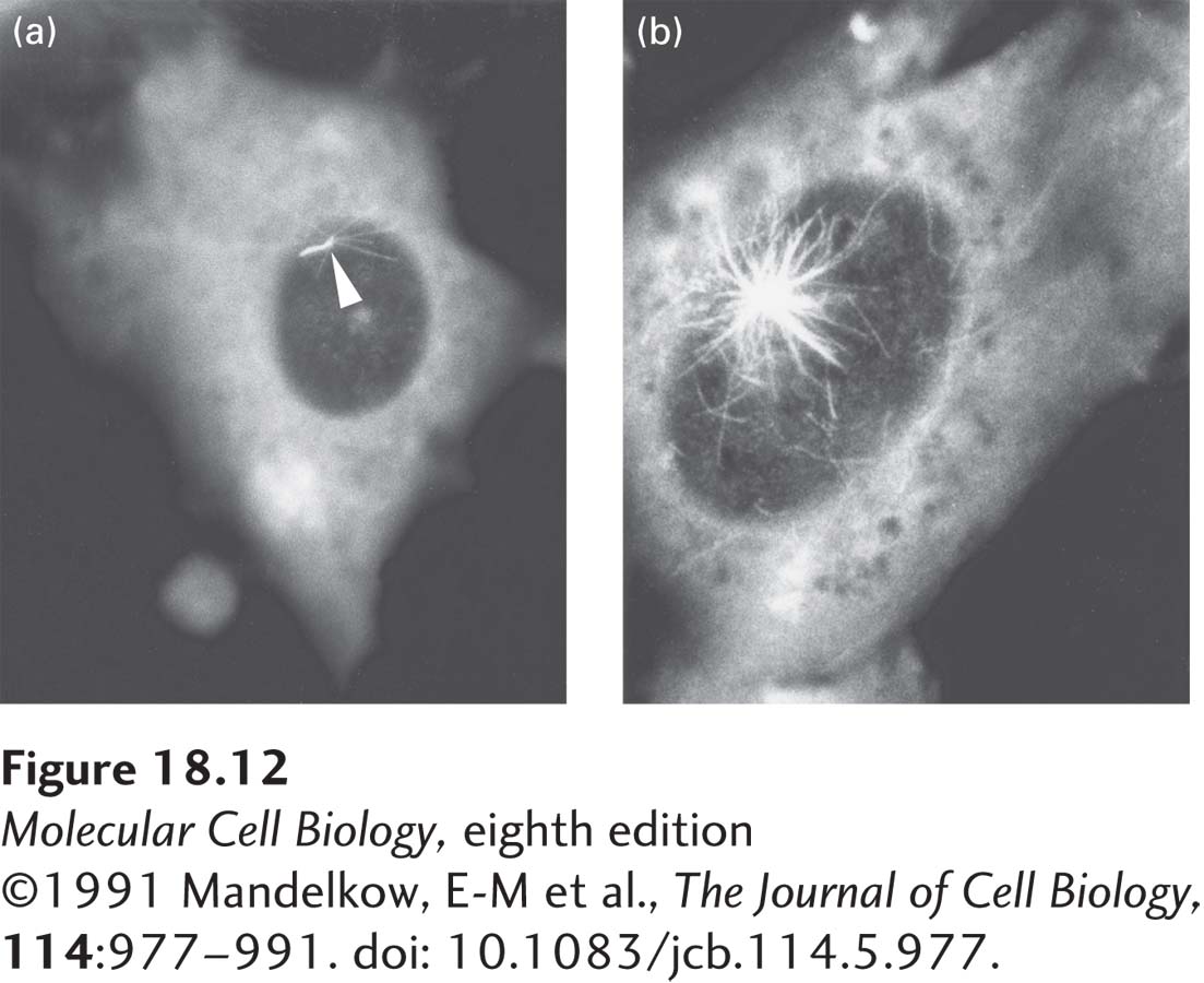
EXPERIMENTAL FIGURE 18-12 Microtubules grow from the MTOC. To investigate the assembly of microtubules in vivo, a cultured fibroblast was treated with colchicine until almost all the cytoplasmic microtubules were disassembled. The cell was then stained with antibodies to tubulin and viewed by immunofluorescence microscopy (a). The colchicine was then washed out to allow the reassembly of microtubules. Panel (b) shows the first stages of reassembly, revealing microtubules growing from the MTOC in the central region above the nucleus (dark areas). Note in panel (a) the remaining primary cilium (arrowhead; discussed in Section 18.5) associated with the centrosome, which is not depolymerized by colchicine treatment under these conditions. Note also the fluorescence from the cytoplasm, which is from unpolymerized αβ-tubulin dimers.
[ ©1991 Mandelkow, E-M et al., The Journal of Cell Biology, 114:977–991. doi: 10.1083/jcb.114.5.977.]