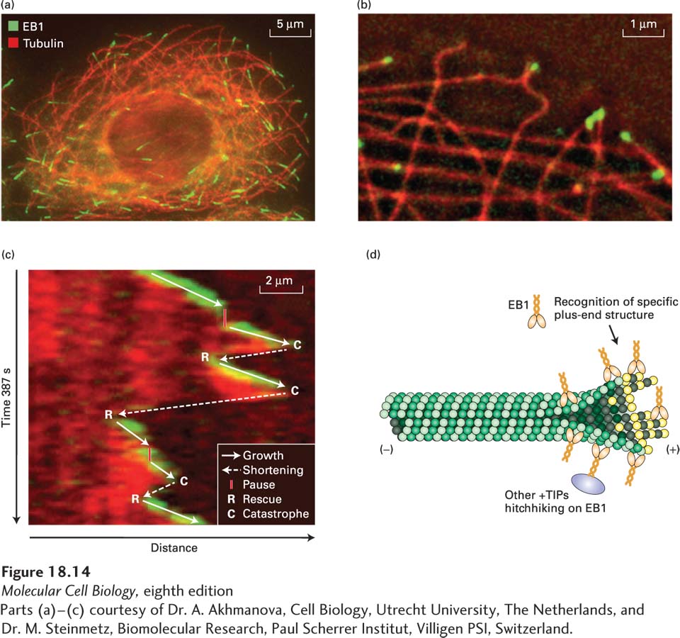
image
EXPERIMENTAL FIGURE 18-14 The +TIP protein EB1 associates dynamically with the (+) ends of microtubules. (a) A cultured cell stained with antibodies to tubulin (red) and the +TIP protein EB1 (green). EB1 is enriched in the region of the microtubule (+) end. (b) Edge of a live cell expressing EB3-GFP (green) and mCherry-α-tubulin (red). EB3, which is closely related to EB1, is found at the ends of some microtubules. (c) EB3-GFP selectively associates with growing microtubules, as seen in this so-called kymograph. In this figure, the dynamics of a single microtubule (red) and EB3 (green) in a live cell like that shown in part (b) is followed by taking the same region from sequential frames of a movie and lining them up top to bottom. At the top, one sees the start of the movie with the microtubule capped by EB3. Moving down the figure, one can track the dynamics of the microtubule over time as it grows and shrinks. When the microtubule grows, it remains capped by EB3. When microtubule growth pauses or the microtubule shrinks, EB3 is no longer associated with the end, but it becomes reassociated when growth resumes. A diagrammatic summary of the microtubule dynamics overlies the kymograph. (d) A possible mechanism for EB1 binding to a growing microtubule and “hitchhiking” by other proteins on EB1.
[Parts (a)–(c) courtesy of Dr. A. Akhmanova, Cell Biology, Utrecht University, The Netherlands, and Dr. M. Steinmetz, Biomolecular Research, Paul Scherrer Institut, Villigen PSI, Switzerland.]