image
EXPERIMENTAL FIGURE 18-17 DIC microscopy demonstrates microtubule-based vesicle transport in vitro. Cytoplasm was squeezed from a squid giant axon with a roller onto a glass coverslip. After buffer containing ATP was added to the preparation, it was viewed by differential interference contrast (DIC) microscopy, and the images were recorded on videotape. In the sequential images shown, the two organelles indicated by open and solid triangles move in opposite directions (indicated by colored arrows) along the same filament, pass each other, and continue in their original directions. Elapsed time in seconds appears at the upper-right corner of each video frame.
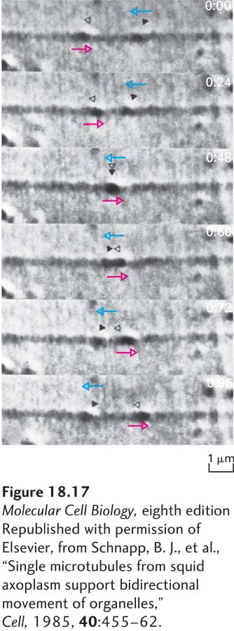
[Republished with permission of Elsevier, from Schnapp, B. J., et al., “Single microtubules from squid axoplasm support bidirectional movement of organelles,” Cell, 1985, 40:455–62.]