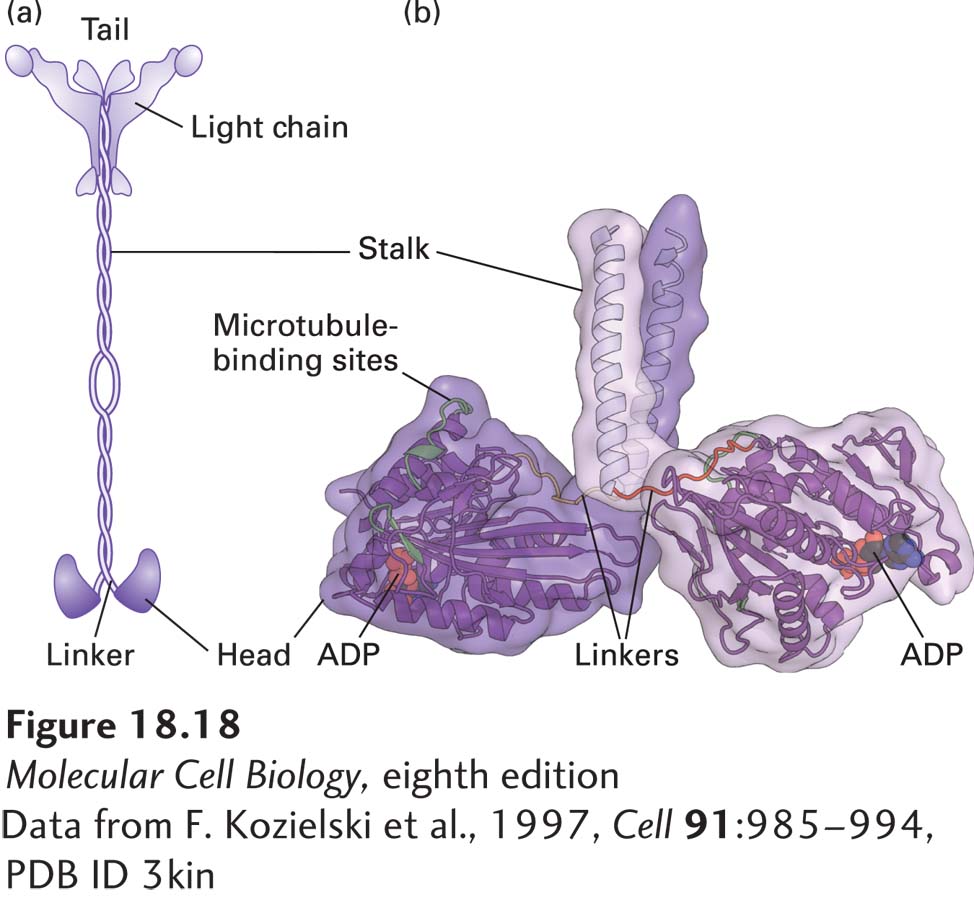
FIGURE 18-18Structure of kinesin-1. (a) Representation of kinesin-1 showing its two intertwined heavy chains, each with a motor domain in the head region. Each head is attached to the coiled-coil stalk by a flexible linker domain. Two light chains associate with the tail of the heavy chain. See R. D. Vale, 2003, Cell112:467. (b) X-ray structure of the kinesin heads with the microtubule-binding and nucleotide-binding sites (containing ADP) indicated, including the linkers and the beginning of the stalk region. See M. Thormahlen et al., 1998, J. Struct. Biol.122:30.
[Data from F. Kozielski et al., 1997, Cell91:985–994, PDB ID 3kin]