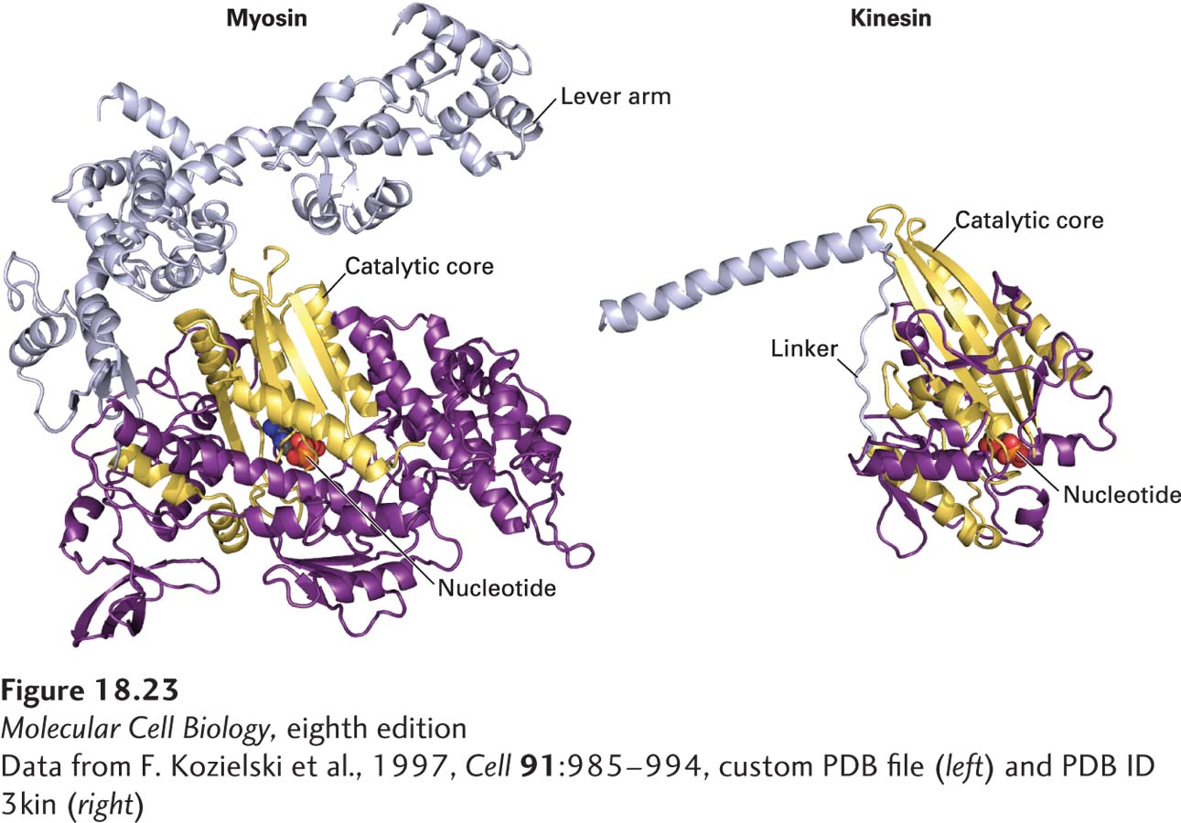
image
FIGURE 18-23 Convergent structural evolution of the ATP-binding cores of myosin and kinesin heads. The common catalytic cores of myosin and kinesin are shown in yellow, the nucleotide in red, and the lever arm (for myosin-II) and linker domain (for kinesin-1) in light purple. See R. D. Vale and R. A. Milligan, 2000, Science 288:88.
[Data from F. Kozielski et al., 1997, Cell 91:985-994, custom PDB file (left) and PDB ID 3kin (right)]