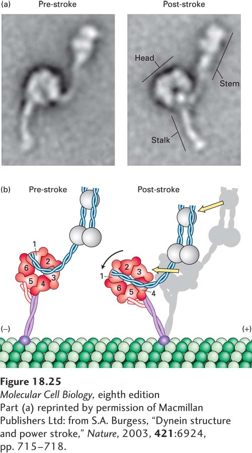
image
FIGURE 18-25 The power stroke of dynein. (a) Multiple images of purified single-headed dynein molecules in their pre-stroke and post-stroke states were recorded in an electron microscope and then averaged. The image at the left shows dynein in the ADP + Pi state, which represents the pre-stroke state, and the image at the right shows it in a nucleotide-free post-stroke state. (b) A comparison of the microscopic images combined with recently acquired structural data shows that the force-generation mechanism involves a change in the position of the linker, which causes a movement of the microtubule-binding stalk. See G. Bhabha et al., 2014, Cell 159:857.
[Part (a) reprinted by permission of Macmillan Publishers Ltd: from S.A. Burgess, “Dynein structure and power stroke,” Nature, 2003, 421:6924, pp. 715-718.