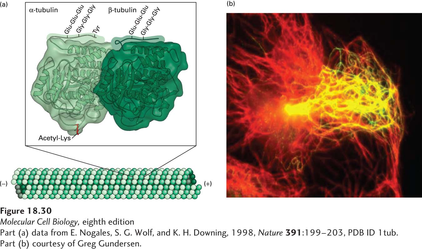
image
EXPERIMENTAL FIGURE 18-30 Post-translational modifications of tubulin affect the stability and function of microtubules. (a) Structure of α- and β-tubulin, showing the sites of lysine acetylation on the inner surface of the microtubule and polyglutamylation, polyglycylation, and detyrosylation on the outer surface. Note that polyglutamylation and polyglycylation are believed to be mutually exclusive, so they would not normally occur at the same time. See J. W. Hamond, D. Cai, and K. J. Verhey, 2008, Curr. Opin. Cell Biol. 20:71–76. (b) Detyrosylated microtubules are preferentially oriented toward the leading edge of a moving cell. A cell migrating toward the right is stained for total microtubules (red) and detyrosylated microtubules (green). The resulting merged image shows the detyrosylated microtubules enriched toward the front of the cell in yellow, which is a combination of red and green.
[Part (a) data from E. Nogales, S. G. Wolf, and K. H. Downing, 1998, Nature 391:199–203, PDB ID 1tub. Part (b) courtesy of Greg Gundersen.]