image
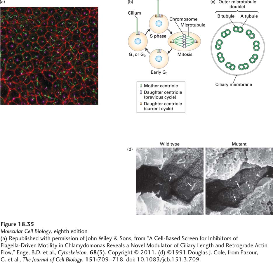
FIGURE 18-35 Many interphase cells contain a nonmotile primary cilium. (a) Fluorescence micrograph of mouse epithelial cells stained with antibodies to acetylated α-tubulin (green), which decorate the primary cilia; to pericentrin (magenta), which decorate the centrosome; and to ZO-1(red), which label the tight junctions that encircle each cell. (b) A diagram depicting how the presence of the primary cilium is tied to the centrioles, one of which serves as its basal body. (c) A diagram depicting a section through a nonmotile primary cilium, showing the lack of the central pair of microtubules and dynein arms typical of motile cilia and flagella. (d) Scanning electron micrographs of epithelial cells of a kidney collecting tubule from a wild-type mouse (left) and a mutant mouse defective in a component of the IFT particles. Arrows point to the primary cilia, which are short stubs in the mutant mouse.
[(a) Republished with permission of John Wiley & Sons, from “A Cell-Based Screen for Inhibitors of Flagella-Driven Motility in Chlamydomonas Reveals a Novel Modulator of Ciliary Length and Retrograde Actin Flow,” Enge, B.D. et al., Cytoskeleton, 68(3). Copyright © 2011. (d) ©1991 Douglas J. Cole, from Pazour, G. et al., The Journal of Cell Biology. 151:709-718. doi: 10.1083/jcb.151.3.709.]