image
FIGURE 18-40 The structure of a mammalian kinetochore. Diagram and electron micrograph of a mammalian kinetochore.
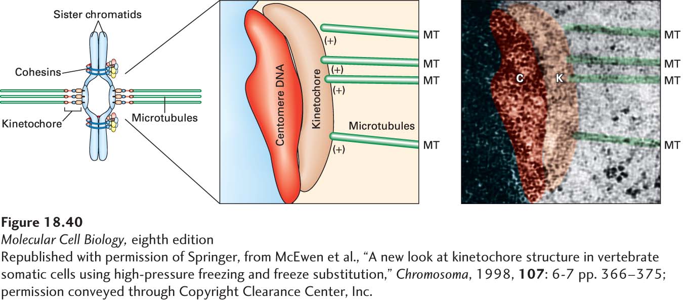
[Republished with permission of Springer, from McEwen et al., “A new look at kinetochore structure in vertebrate somatic cells using high-pressure freezing and freeze substitution,” Chromosoma, 1998, 107: 6-7 pp. 366-375; permission conveyed through Copyright Clearance Center, Inc.]