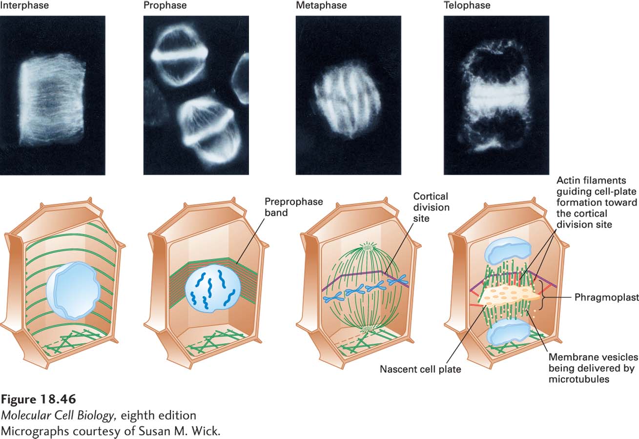
image
FIGURE 18-46 Mitosis in a flowering plant cell. Immunofluorescence micrographs (top) and corresponding diagrams (bottom) showing arrangement of microtubules in interphase and mitotic plant cells. A cortical array of microtubules girdles a cell during interphase. As the cell enters prophase, the microtubules (green), together with actin filaments (red), assemble under the cell cortex into a preprophase band, which marks the future cortical division site. As the cell enters prometaphase and metaphase, a spindle similar to that seen in animal cells forms. However, due to the cell wall, cytokinesis in plant cells is very different from that in in animal cells. Microtubules deliver vesicles whose membranes are used to assemble a membrane network called a phragmoplast, whose organization is defined by actin filaments linked to the cortical division site. Eventually, the phragmoplast becomes part of the plasma membranes of the two daughter cells. Enzymes secreted from the vesicles then build a cell wall between the two daughter cells. See G. Jürgens, 2005, Annu. Rev. Plant Biol. 56:281–299
[Micrographs courtesy of Susan M. Wick.]