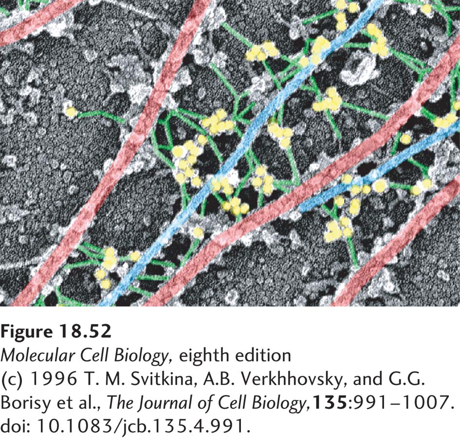
image
EXPERIMENTAL FIGURE 18-52 Gold-labeled antibody identifies plectin cross-links between intermediate filaments and microtubules. In this immunoelectron micrograph of a fibroblast cell, microtubules are highlighted in red, intermediate filaments in blue, and the short connecting fibers between them in green. Staining with gold-labeled antibodies to plectin (yellow) reveals that these connecting fibers contain plectin.
[(c) 1996 T. M. Svitkina, A.B. Verkhhovsky, and G.G. Borisy et al., The Journal of Cell Biology, 135:991–1007. doi: 10.1083/jcb.135.4.991.]