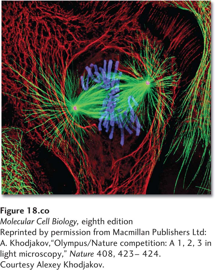
Newt lung cell in mitosis stained for centrosomes (magenta), microtubules (green), chromosomes (blue), and keratin intermediate filaments (red).
[Reprinted by permission from Macmillan Publishers Ltd: A. Khodjakov, “Olympus/Nature competition: A 1, 2, 3 in light microscopy,” Nature 408, 423-