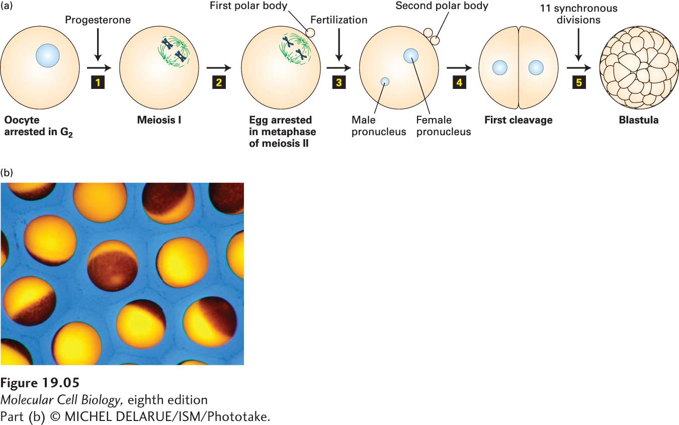
image
FIGURE 19-5 Progesterone stimulates maturation of Xenopus oocytes. (a) Step 1: Progesterone treatment of G2-arrested Xenopus oocytes surgically removed from the ovary of an adult female causes the oocytes to enter meiosis I. Two pairs of synapsed homologous chromosomes (blue) connected to meiotic spindle microtubules (green) are shown schematically to represent cells in metaphase of meiosis I. Step 2: Segregation of homologous chromosomes and a highly asymmetric cell division expels half the chromosomes into a small cell called the first polar body. The oocyte immediately commences meiosis II and arrests in metaphase II to yield an egg. Two chromosomes connected to spindle microtubules are shown schematically to represent egg cells arrested in metaphase of meiosis II. Step 3: Fertilization by sperm releases eggs from their metaphase arrest, allowing them to proceed through anaphase of meiosis II and undergo a second highly asymmetric cell division that expels one chromatid of each chromosome into a second polar body. The resulting haploid female pronucleus fuses with the haploid sperm pronucleus to produce a diploid zygote. Step 4: The zygote undergoes DNA replication and the first mitosis. Step 5: The first mitosis is followed by 11 more synchronous divisions to form a blastula. (b) Micrograph of Xenopus eggs.
[Part (b) © MICHEL DELARUE/ISM/Phototake.]