image
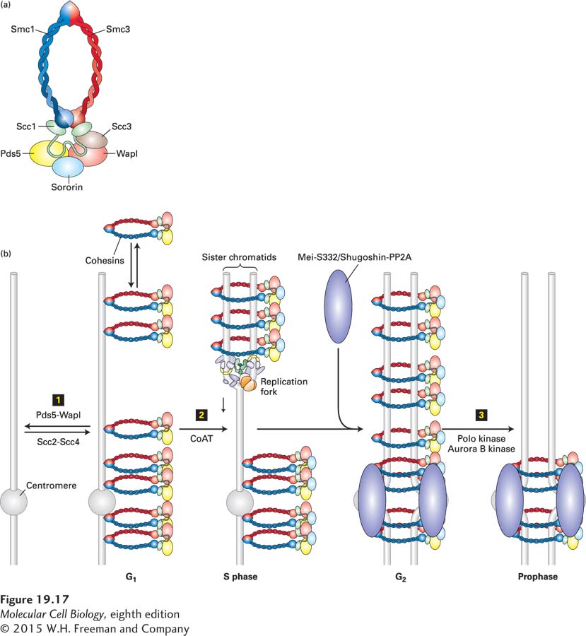
FIGURE 19-17 Model for establishment of cohesin linkage of sister chromatids. Cohesin complexes form rings that link sister chromatids by embracing the two sister DNA molecules. (a) Schematic structure of the cohesin complex. (b) Mechanism whereby cohesins are loaded onto DNA and acquire their cohesive properties. Step 1: Cohesins are loaded onto chromosomes during G1 by the cohesin-loading complex Scc2-Scc4, but they do not possess cohesive properties (indicated as cohesins laterally associated with chromosomes). In this state, cohesins are dynamic and can dissociate from the DNA with the help of the Pds5-Wapl complex, which associates with cohesins. Step 2: Concomitant with DNA replication, closely behind the replication fork, cohesins are converted into cohesive molecules, able to hold sister chromatids together (indicated as cohesin rings encircling the replicated sister chromatids), through acetylation of Smc3 by cohesin acetyltransferases (CoATs). Acetylation is accompanied by the binding of sororin to cohesin, which helps stabilize cohesins on chromosomes. During G2, sister chromatids are replicated and linked along their entire length by cohesins. During this time, the Mei-S332/Shugoshin proteins recruit the protein phosphatase 2A (PP2A) to centromeric regions. Step 3: In vertebrate cells, cohesins are released from chromosome arms during prophase and early metaphase by the action of the Pds5-Wapl complex and phosphorylation of cohesins by Polo kinase and Aurora B kinase. By the end of metaphase, cohesins are retained only in the region of the centromere, where Mei-S332/Shugoshin prevents cohesin phosphorylation, and hence dissociation, by recruiting PP2A.