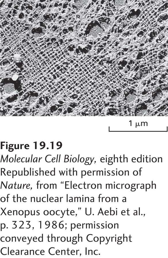
Figure 19- h-
[Republished with permission of Nature, from “Electron micrograph of the nuclear lamina from a Xenopus oocyte,” U. Aebi et al., p. 323, 1986; permission conveyed through Copyright Clearance Center, Inc.]