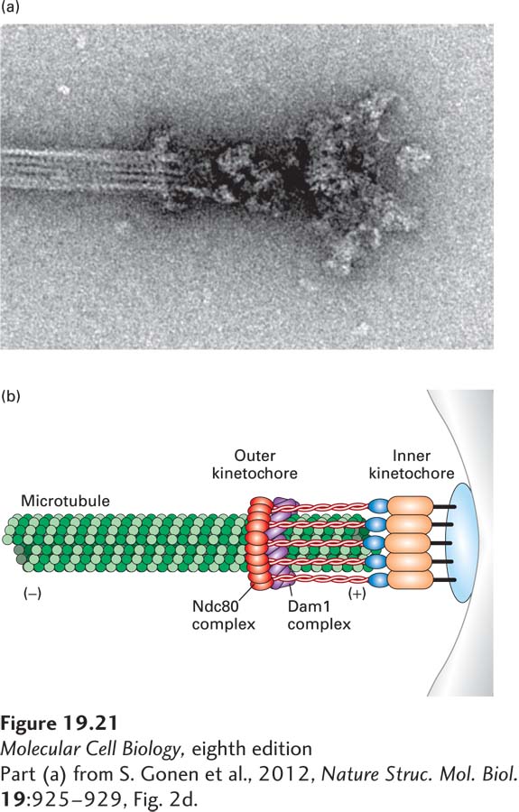
image
FIGURE 19-21 Electron micrograph of purified yeast kinetochores bound to taxol-stabilized microtubules. (a) End-on attachment of a yeast kinetochore at a microtubule. (b) A cartoon schematizing the key features of the image. The ring structure embracing the microtubule most likely represents the outer kinetochore Dam1 complex and part of the Ndt80 complex, also an outer kinetochore component. The globular structure at the end of the complex most likely reflects the inner kinetochore and protein complexes that link the inner kinetochore to the outer kinetochore.
[Part (a) from S. Gonen et al., 2012, Nature Struc. Mol. Biol. 19:925-929, Fig. 2d.]