image
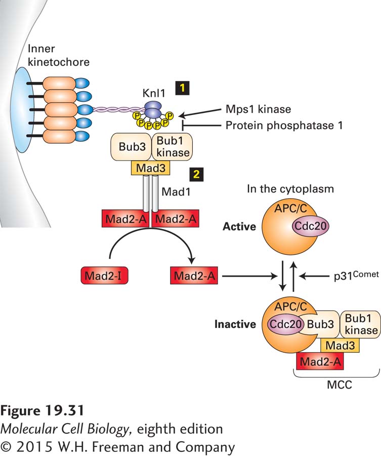
FIGURE 19-31 The spindle assembly checkpoint pathway. The spindle assembly checkpoint pathway is active until every single kinetochore has attached properly to spindle microtubules. When a kinetochore is unattached, the outer kinetochore component Knl1 is phosphorylated by the checkpoint kinase Mps1 step 1. Phosphorylated Knl1 then binds the checkpoint kinase Bub1-Bub3 and the checkpoint protein Mad3. These three proteins, in turn, recruit the Mad1-Mad2 complex to the kinetochore. The Mad1-Mad2 complex bound to a kinetochore is in the active form (shown as Mad2-A; step 2 ) and has the ability to convert inactive Mad2 (Mad2-I) in the cytoplasm into active Mad2 that is able to bind APC/CCdc20 and inhibit it. Complete inhibition of APC/CCdc20 requires the recruitment of the checkpoint factors Bub1-Bub3 and Mad3 into the complex. Together, these proteins form the mitotic checkpoint complex (MCC) that prevents APC/CCdc20 from recognizing and ubiquitinylating its substrates. Silencing of the spindle assembly checkpoint pathway occurs once all kinetochores have attached to microtubules in a tension-generating manner. Protein phosphatase 1 then dephosphorylates Knl1, thereby eliminating checkpoint protein binding sites at kinetochores. In addition, p31comet disassembles the MCC. See E. Foley and T. Kapoor, 2013, Nature Rev. Mol. Cell Biol. 14:25.