image
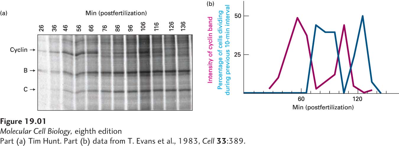
FIGURE 1 Autoradiography permits the detection of cyclical synthesis and destruction of mitotic cyclin in sea urchin embryos. A suspension of sea urchin eggs was synchronously fertilized by the addition of sea urchin sperm, and 35S-methionine was added. At 10-minute intervals beginning 26 minutes after fertilization, samples were taken for protein analysis on an SDS-polyacrylamide gel and for detection of cell cleavage by microscopy. (a) Autoradiogram of the SDS gel showing samples removed at each time point. The bands for most proteins, such as B and C, continuously increased in intensity. In contrast, the band for cyclin suddenly decreased in intensity at 76 minutes after fertilization and then began increasing again at 86 minutes. The cyclin band peaked again at 106 minutes and decreased again at 126 minutes. (b) Plot of the intensity of the cyclin band (red line) and the percentage of cells that had undergone cleavage during the previous 10-minute interval (cyan line). Note that the amount of cyclin fell precipitously just before cell cleavage.
[Part (a) Tim Hunt. Part (b) data from T. Evans et al., 1983, Cell 33:389.]