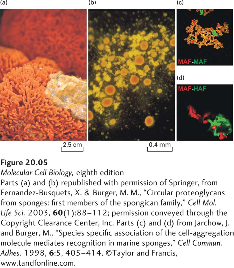
EXPERIMENTAL FIGURE 20- 5 Mechanically separated marine sponges reassemble through species- specific homotypic cell adhesion. (a) Two intact sponges, Microciona prolifera (orange) and Halichondria panicea (yellow), growing in the wild. (b) After mechanical disruption and mixing of the individual cells from the two sponge species, their individual cells were allowed to reassociate for about 30 minutes with gentle stirring. The cells aggregated with species- specific homotypic adhesion, forming clumps of M. prolifera cells (orange) and H. panicea cells (yellow). (c) and (d) Red or green fluorescently labeled beads were coated with the proteoglycan aggregation factor (AF) from the ECM of either M. prolifera (MAF) or H. panicea (HAF). (c) When beads of both colors were coated with MAF, they all aggregated together, forming yellow aggregates (combination of red and green). (d) MAF (red) and HAF (green) coated beads do not readily form mixed aggregates, but rather assemble into distinct clumps held together by homotypic adhesion. (Magnification 40×.)
[Parts (a) and (b) republished with permission of Springer, from Fernandez- Busquets, X. & Burger, M. M., “Circular proteoglycans from sponges: first members of the spongican family,” Cell Mol. Life Sci. 2003, 60(1):88– 112; permission conveyed through the Copyright Clearance Center, Inc. Parts (c) and (d) from Jarchow, J. and Burger, M., “Species- specific association of the cell- aggregation molecule mediates recognition in marine sponges,” Cell Commun. Adhes. 1998, 6:5, 405– 414, ©Taylor and Francis, www.tandfonline.com.]
[Leave] [Close]