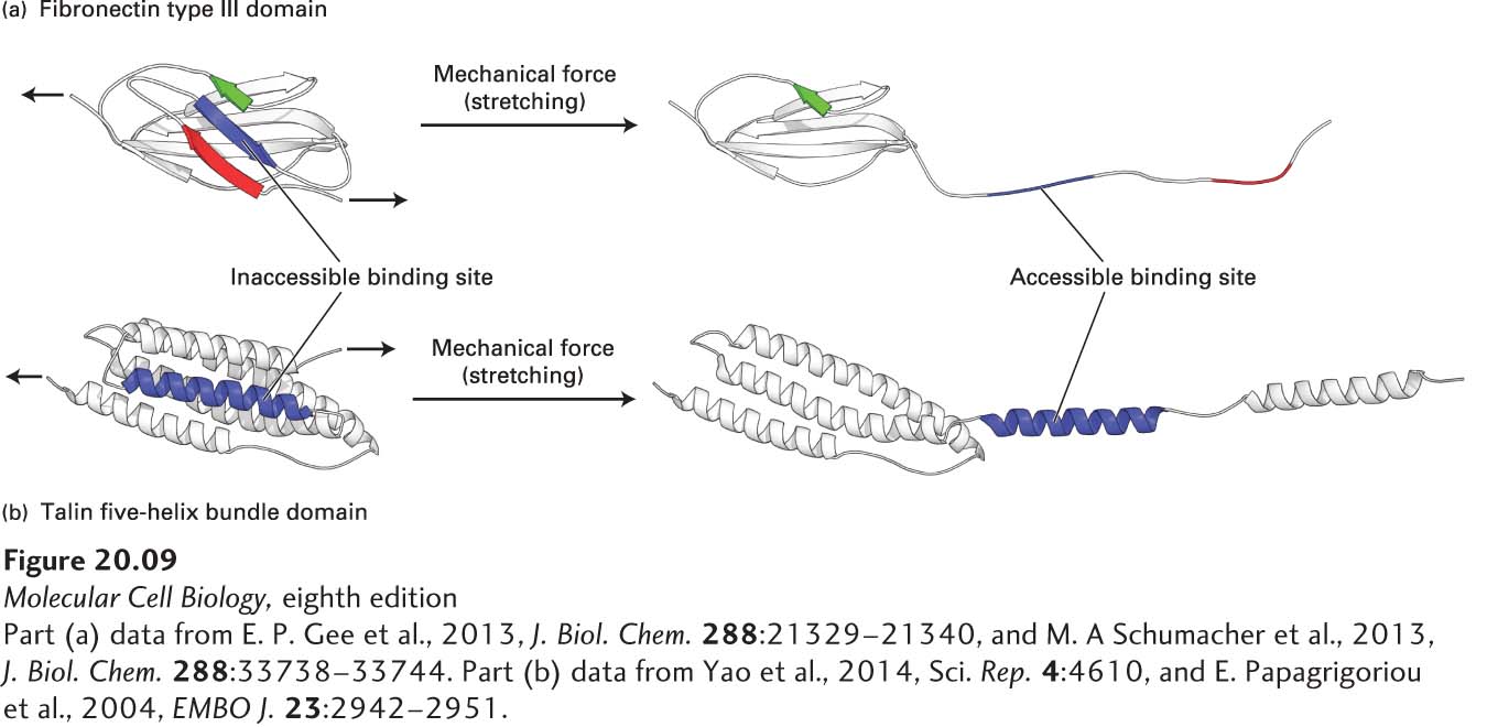
FIGURE 20- 9 Models of Domains in Mechanosensor Proteins Responding to Mechanical Forces. (a) Hypothetical model of the partial unfolding of a fibronectin type III domain in the ECM molecule fibronectin when that protein is subjected to mechanical force. Mechanical force generated within the cell by actin movement and mechanotransduced via multiple integrin adhesion receptors bound to the extracellular dimeric fibronectin can partially unfold the fibronectin. The unfolding is thought to expose a putative, previously hidden (cryptic) binding site on fibronectin (blue segment) that has the potential to form β sheets with other fibronectin molecules, recruiting them to form fibronectin fibrils, and thus helping assemble the ECM. (b) Hypothetical model of the partial unfolding of a domain (the R1 five- helix bundle) in the intracellular integrin adapter protein talin when it is subjected to mechanical stretching force. This force is generated by actin, which can bind to and pull on the C- terminus of talin while talin’s N- terminus is bound to the cytoplasmic tail of integrin’s β subunit. The unfolding is thought to expose this domain’s otherwise cryptic α-helical vinculin binding site (blue). Vinculin, an actin- binding protein (see Figure 20- 14d ), can then bind to the integrin- talin complex via the exposed site and in turn bind to actin, thus promoting the assembly of multiple actin fibers. The assembly of actin fibers indirectly linked by adapters to integrins strengthens integrin- mediated adhesion and helps to build focal adhesions.
[Part (a) data from E. P. Gee et al., 2013, J. Biol. Chem. 288:21329– 21340, and M. A Schumacher et al., 2013, J. Biol. Chem. 288:33738– 33744. Part (b) data from Yao et al., 2014, Sci. Rep. 4:4610, and E. Papagrigoriou et al., 2004, EMBO J. 23:2942– 2951.]
[Leave] [Close]