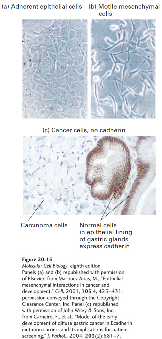
EXPERIMENTAL FIGURE 20- 15 E- cadherin activity is lost during the epithelial- to- mesenchymal transition and during cancer progression. A protein called Snail that suppresses the expression of E- cadherin is associated with the epithelial- to- mesenchymal transition (EMT). (a) Normal epithelial MDCK cells grown in culture. (b) Expression of the snail gene in MDCK cells causes them to undergo an EMT. (c) Distribution of E- cadherin detected by immunohistochemical staining (dark brown) in thin sections of tissue from a patient with hereditary diffuse gastric cancer. E- cadherin is seen at the intercellular borders of normal stomach gastric gland epithelial cells (right); no E- cadherin is seen at the borders of underlying invasive carcinoma cells.
[Panels (a) and (b) republished with permission of Elsevier, from Martinez Arias, M., “Epithelial mesenchymal interactions in cancer and development,” Cell, 2001, 105:4, 425– 431; permission conveyed through the Copyright Clearance Center, Inc. Panel (c) republished with permission of John Wiley & Sons, Inc., from Carneiro, F., et al., “Model of the early development of diffuse gastric cancer in Ecadherin mutation carriers and its implications for patient screening,” J. Pathol., 2004, 203(2):681– 7.]
[Leave] [Close]