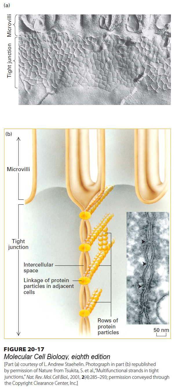
FIGURE 20- 17 Tight junctions. (a) Freeze- fracture preparation of tight junction zone between two intestinal epithelial cells. The fracture plane passes through the plasma membrane of one of the two adjacent cells (see also Figure 20- 11 ). A honeycomb- like network of ridges and grooves below the microvilli constitutes the tight- junction zone. (b) Schematic drawing shows how a tight junction might be formed by the linkage of rows of protein particles in adjacent cells. In the inset micrograph of an ultrathin sectional view of a tight junction, the adjacent cells can be seen in close contact where the rows of proteins interact. See L. A. Staehelin and B. E. Hull, 1978, Sci. Am. 238:140, and D. Goodenough, 1999, P. Natl. Acad. Sci. USA 96:319.
[Part (a) courtesy of L. Andrew Staehelin. Photograph in part (b) republished by permission of Nature, from Tsukita, S. et al., “Multifunctional strands in tight junctions,” Nat. Rev. Mol. Cell Biol., 2001, 2(4):285– 293; permission conveyed through the Copyright Clearance Center, Inc.]
[Leave] [Close]