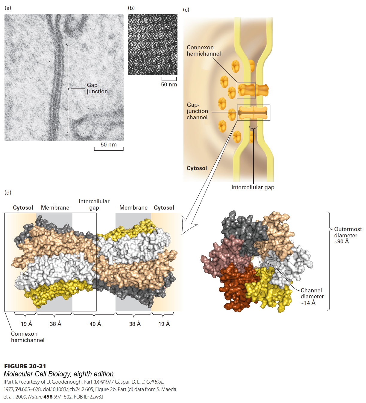
FIGURE 20- 21 Gap junctions. (a) In this thin section through a gap junction connecting two mouse liver cells, the two plasma membranes are closely associated for a distance of several hundred nanometers, separated by a “gap” of 2– 3 nm. (b) Numerous roughly hexagonal particles are visible in this perpendicular view of the cytosolic face of a region of plasma membrane enriched in gap junctions. Each hexagonal particle aligns with a similar particle on an adjacent cell, forming a channel connecting the two cells. (c) Schematic model of a gap junction connecting two plasma membranes. Both membranes contain connexon hemichannels, cylinders of six dumbbell- shaped connexin molecules. Two connexons join in the gap between the cells to form a gap- junction channel, 1.4– 2.0 nm in diameter, that connects the cytosols of the two cells. (d) Structure of recombinant human Cx26 gap junction as determined by x- ray crystallography (3.5- Å resolution). Left: Space- filling model of a side view of the complete structure of two attached connexons oriented as in part (c). Each of the six connexins that comprise a connexon has four transmembrane helices and is shown in a distinct color. The structures of the loops connecting the transmembrane helices are not well defined and not shown. Right: View from the cytosol perpendicular to the membrane bilayers, looking down on the connexon with its central pore. The diameter of the pore’s channel is ~14 Å, and it is lined by many polar/charged amino acids. See S. Nakagawa et al., 2010, Curr. Opin. Struct. Biol. 20(4):423– 430.
[Part (a) courtesy of D. Goodenough. Part (b) ©1977 Caspar, D. L., J. Cell Biol., 1977, 74:605– 628. doi:10:1083/jcb.74.2.605; Figure 2b. Part (d) data from S. Maeda et al., 2009, Nature 458:597– 602, PDB ID 2zw3.]
[Leave] [Close]